A team of Kiwi scientists is designing a 3D scanner that could revolutionise how diseases like cancer are diagnosed.
The invention of the modern X-Ray was a medical milestone. It's hard to imagine a world where broken bones were once detected through physical examination. And now, a team of Kiwi scientists is designing a 3D scanner that could revolutionise x-ray technology once more, helping to detect diseases like cancer in a totally new way.
The joint initiative between the University of Canterbury, University of Otago, and MARS Bioimaging Ltd, known as the MARS Programme, is producing a spectral molecular scanner which provides 3D colour images of objects inside the body such as bone, soft tissue, and artificial joints. It's just been backed by the Ministry of Business, Innovation and Employment (MBIE) with "gold status" for research excellence.
One of the lead researchers, University of Canterbury Professor Anthony Butler, says the research team is at the forefront of this new technology that will deliver significant health benefits.
“This spectral molecular imaging technology really is the next big medical imaging innovation, and these 3D images will provide clinicians with information that is currently not possible in CT, MRI or PET scans,” says Professor Butler.
“This technology really is the next big medical imaging innovation, and these 3D images will provide clinicians with information that is currently not possible in CT, MRI or PET scans."
“The implications of this research will be huge for the medical profession. The capability of this scanner will enable greater diagnosis and monitoring of many diseases, and will lead to better outcomes for patients – particularly in stroke prevention, joint replacement and cancer management.
“The technology also has significant financial benefits, with the potential to add more than $50 million per year to the New Zealand economy once the manufactured products arising from this research are in regular use in hospitals.”
The team’s main goal is for MARS Bioimaging Ltd to market MARS scanners developed by the University of Canterbury, where proof of concept trials have been undertaken at Otago University Christchurch. The initial market is a range of researchers from many institutes. The research has also been supported by the MedTech Centre of Research Excellence and GE Healthcare. GE is providing a top of the line CT scanner to the MARS team to accelerate the research. As an another example of the interdisciplinary nature of the MARS programme, the GE scanner will be installed in the animal facilities at Lincoln University.
The team will also explore potential applications in other industries such as border security, forestry, agriculture and mining, Professor Butler says.
UC Professor Phil Butler, science leader at the start of this MARS project and now CEO of MARS Bioimaging Ltd, says: “Medical innovations have high commercial value and high social impact, but long lead times. The strong support of MBIE has been essential over the duration of this project, a project that is just beginning to break into the pre-clinical research market in the US, India and China.”
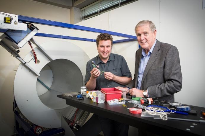
Main photo: Professor Anthony Butler (left) and his father Professor Phil Butler have built the MARS spectral (colour) CT scanner, and demonstrated that the colour information can give novel functional information and novel molecular information. Photo credit: University of Canterbury.